Introduction
Amelogenesis imperfecta (ai) is a hereditary disorder that affects enamel on primary and permanent teeth. It is reported to have an incidence of one person in every 14,000[1],[2]. Although amelogenesis imperfecta has been categorized into 4 broad groups primarily based on phenotype—hypoplastic, hypocalcified, hypomaturation, and hypomaturation-hypoplastic - at least 15 subtypes of ai exist when phenotype and mode of inheritance are considered [2],[4]. According to the literature, ai patients, regardless of subtype, have similar oral complications: teeth sensitivity, poor dental aesthetics, and decreased occlusal vertical dimension. Other dental anomalies associated with ai include, but are not limited to, multiple impacted teeth, congenitally missing teeth, open occlusal relationship, and taurodontism [3].
Historically, patients with ai have been treated with multiple extractions and the fabrication of complete dentures [5],[6],[7],[8]. Recently, several studies have illustrated the use of stainless steel crowns, adhesive casting, over denture, porcelain veneers, ceramics, and composite resin veneers to restore dentitions mutilated by severe attrition[9],[10],[11]. Besides, the advances in the field of aesthetic dentistry, especially in bonding to dentin, help practitioners to restore function and aesthetics to an acceptable level.
This clinical report describes treatment of a 17-year-old girl patient diagnosed with hypomature amelogenesis imperfecta by using composite resin veneers, post and core and ceramic crowns.
Case Report
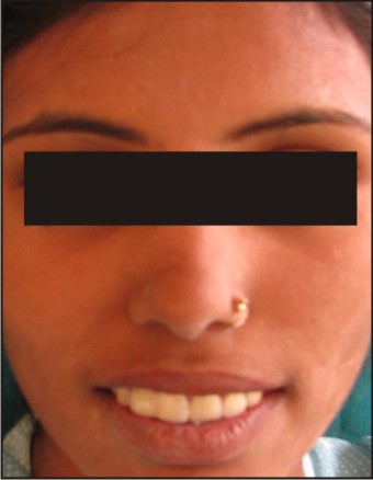
A 17-year-old girl was referred to the department of prosthodontics including crown and bridge, for examination, evaluation and treatment of gross discolouration, fractures and considerable sensitivity of her teeth (Fig 1).
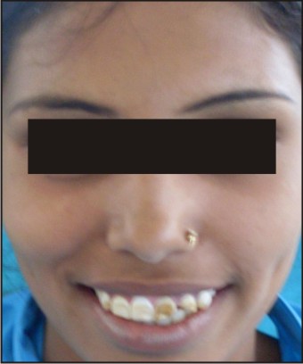 | Fig 1 : Before Treatment
 |
A detailed medical, dental, and social history was obtained. She was both self-conscious and unhappy as regard to the appearance of her teeth.
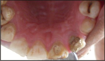
Clinical examination revealed that the enamel layer was very thin and brown (Pigmented), the cuspal structure was completely absent in the occlusal portion of the molars and enamel pit defects (pigmented stains deep) were present in the anterior teeth (Fig 2). However, the clinical appearance of cervical and approximal enamel seemed to be normal. It was thought that the patient likely suffered from a hypomature type of ai. The exposed dentin was hypersensitive. Radiographic examination of the patient revealed deep carious lesions in the maxillary, and the mandibular molars and fractured maxillary left anterior incisors and canine (Fig 3).
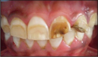 | Fig 2 : Before Treatment (Intraoraly)
|
 | Fig 3 : Opg
|
A treatment plan was developed with the following aims: to reduce the reported sensitivity of the teeth, to restore fractures and to improve the aesthetics. The patient was informed of the diagnosis and all treatment plans were discussed with her and his parents.
Steps of treatment followed-
First, root canal treatment were done for maxillary anterior left incisors, followed with custom made post placement in maxillary left lateral incisor (Fig 4).
 | Fig 4 : Root Canal Treatment Done On Left Lateral & Central Incisor Followed By Custom Made Post Placement On Left Lateral Incisor
|
Then ,tooth preparation was done for porcelain fused to metal crowns on left maxillary incisors and canine and futher on right maxillary incisors and canine for composite veneers (Fig 5).
 | Fig 5 : Tooth Preparation Done On Left Incisors And Canine For Placement Of Porcelain Fused To Metal Crowns
|
Metal try-in done for ceramic crowns and shade matching was then done .
Finally composite veneering was done on right maxillary anteriors and porcelain fused to metal crowns on left maxillary anteriors.(Fig 6)
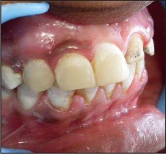 | Fig 6 : Composite Veneering Done On Right Incisors And Canine
|
The patient was recalled at 2-month intervals. On recall clinical and radiographic examination revealed no pathology associated with the rehabilitation, and the patient’s aesthetic and functional expectations were satisfied (Fig 7, 8).
 | Fig 7 : After Treatment
|
 | Fig 8 : After Treatment (Intraoraly)
|
Discussion
The term “amelogenesis imperfecta” describes a diverse group of hereditary conditions primarily affecting the quality and/or quantity of dental enamel. The affected teeth show a soft enamel of normal thickness that chips and wears easily and has a radiodensity similar to that of dentin[2],[3]. The results of clinical and radiographic evaluations indicated that the patient in the present case had hypomaturation form ai. All the teeth are misshapen, and spotted. Occlusion and vertical opening are rapidly affected by attrition. The insufficiency of the enamel makes the teeth extremely sensitive to contact and thermal stimuli. These problems combine to make early diagnosis essential and immediate treatment a necessity, even for the youngest patients[4],[6].
In some types of ai, the patient’s enamel not only is thin but also may display abnormal mineral content. The axial surfaces may be chalky, weak, and highly susceptible to carious breakdown. Such teeth require complete coverage with preformed crowns until precision cast crowns can be provided in the patient’s late teens or early adulthood.
A complex case will challenge any direct composite system. A quality aesthetic direct composite system must contain a wide variety of shades and include multiple opacities to ensure the restorations will mimic natural dentition. As teeth are not monochromatic and the colors come from within, the color must be developed from the inside out [7],[8],[9]. The polish of a quality direct composite system should provide high gloss and should be maintained in the oral environment.
Walter[8] applied composite resin restorations for anterior teeth to 10-year-old child with ai as provisional treatment. Similarly, in the present case the treatment was completed with direct composite resin. Although this case could have been treated with indirect restorations, direct resin treatment was chosen to preserve tooth structure and for financial reasons. When all the teeth had been restored direct composite resin, the patient’ aesthetic complaints disappeared completely, and normal eating habits were established. Temporary restorations provide satisfactory aesthetic and protective features with minimum preparation, made difficult by crown height and lack of enamel in untreated amelogenesis imperfecta, to be performed under excellent conditions. This temporary treatment allows the normal development of the dentition to continue unimpaired and, last but not least, is affordable for the parents.
It is very important in that it prevents the development of psychological problems arising from the appearance of teeth affected by amelogenesis imperfecta[11]. The shy and taciturn 17-year-old girl first brought forcefully to the department, became a willing and talkative patient. The psychological transformation was spectacular, as was the physiological change, evidenced by a weight gain within weeks.
Conclusion
This clinical report describes the oral rehabilitation of a 17-year-old girl patient affected by hypomature amelogenesis imperfecta. The treatment plan for cases of ai is related to many factors: the age of the patient, the socioeconomic status of the patient, the type and severity of the disorder, and the intraoral situation at the time the treatment is planned. The rehabilitation included anterior composite resins veneers and ceramic crowns to eliminate tooth sensitivity, improve the aesthetics and restore function.
References
1. C. J. Witkop jr. Amelogenesis imperfecta, dentinogenesis imperfecta and dentin dysplasia revisited: problems in classification. Journal of oral pathology 1988;17:547–553.
2. W. K. Seow.clinical diagnosis and management strategies of amelogenesis imperfecta variants. Pediatric dentistry 1993;15:384–393.
3. P. J. M. Crawford, m. Aldred, and a. Bloch-zupan. Amelogenesis imperfecta. Orphanet journal of rare diseases 2007; 2:17.
4. A. Sengün and f. Özer. Restoring function and esthetics in a patient with amelogenesis imperfecta: a case report.quintessence international 2002;33:199–204.
5. K. M. S. Ayers, b. K. Drummond, w. J. Harding, s. G. Salis, p. N. Liston. Amelogenesis imperfecta—multidisciplinary management from eruption to adulthood. Review and case report. New zealand dental journal 2004;100:101–104
6. D. J. Lamb .the treatment of amelogenesis imperfect the journal of prosthetic dentistry 1976 ;36:286–291.
7. R.-r. A. Patel, s. Hovijitra, a. H. Kafrawy, d. Bixler. X-linked (recessive) hypomaturation amelogenesis imperfecta: a prosthodontic, genetic, and histopathologic report. Journal of prosthetic dentistry 1991;66:398–402.
8. B. Walter. Prosthetic rehabilitation of a case of total amelogenesis imperfecta,” actualites odonto-stomatologiques 1991;45:213–226.
9. K. Gökçe, c. Canpolat, e. Özel. Restoring function and esthetics in a patient with amelogenesis imperfecta: a case report. Journal of contemporary dental practice 2007;8 95–101.
10. R. Bedi.the management of children with amelogenesis imperfecta. Restorative dentistry 1989;5:28–34.
11. I. C. Mackie , a. S. Blinkhorn. Amelogenesis imperfecta: early interception to prevent attrition. Dental update 1991;18:79–80. |